bioavailability
If your supplement has high bioavailability, then you're getting what you paid for.

Clinical Studies

Written by MacuHealth
Reviewed by Jim Stringham, Ph.D.

The nutrients included in high-quality supplements offer plenty of health benefits for the human body and fill the nutritional gaps missing from the modern diet. But not all of them are created equal.

“We confirm that a number of commercially available carotenoid food supplements do not achieve their label claim,” explained Dr. David Phelan, Professor John Nolan and Dr. Alfonso Prado-Cabrero in their study on supplements in the journal Nutrients.1

A nutritional supplement’s effectiveness depends on absorption inside the body. But if it offers little to no bioavailability or your body has a difficult time absorbing its nutrients, then it’s not doing you any good.

What is bioavailability, and why is it so important? Bioavailability is effectively absorption – the fraction of active ingredients absorbed into the body’s circulation. It’s dependent on many factors. Knowing if a supplement has high bioavailability is crucial for a nutrient to be effective. Bioavailability also ensures that consumers are getting what they’re paying for. We’ll look at how bioavailability is measured and how supplements retain their potency.

How Does Bioavailability Work?

We absorb the ingredients we ingest in different ways. Some nutrients, such as carbohydrates and proteins, are easily processed into our bloodstream. But vitamins, minerals, antioxidants and other nutrients are more complicated, making it more difficult for our bodies to break them down and absorb them.

For example, a medication injected directly into the bloodstream should have 100 percent bioavailability. But an orally taken drug or supplement must pass through several barriers inside your body, such as your intestinal wall and liver. Some nutrients, such as Vitamin C, are water-soluble, meaning they’re absorbed when taken with water. Other nutrients, such as Vitamin E or carotenoids, are lipid-soluble, meaning that a small amount of fat is necessary for their transport across the intestinal wall into the bloodstream.

With orally ingested nutritional supplements, the fraction received by the body is usually measured with a blood draw. It won’t give you the exact fraction absorbed (higher is better), allowing for comparison among supplements and significant differences between them.

What Can Alter Bioavailability?

Though a supplement’s label may claim a certain amount of an active ingredient, the truth is that due to external factors, low-quality formulations and cost-saving techniques, it might be significantly lower. Factors that can alter the bioavailability of nutrients in supplements include the molecular structure of the active ingredients, the encapsulation process and your body’s own response.

Supplement's Molecular Structure and Formulation

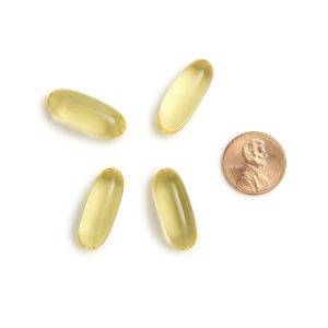
The molecular structure and formulation of ingredients matter. For example, fish oil has re-esterified triglyceride and ethyl ester forms. While the latter is significantly cheaper, it’s much harder to absorb because of its synthetic form. The body recognizes re-esterified triglyceride fish oils, allowing them to be more easily absorbed.

Supplement's Encapsulation Method

Supplements are encapsulated in many forms: powder, softgel, liquid, etc. Powder-filled capsules tend to have a higher chance of degrading and losing bioavailability due to their clear interlock, making them more susceptible to light and oxidation. On the flipside, oil-filled softgels have airtight closing with more protection from heat, light, and oxygen – factors that can significantly degrade nutrients.

Body's Metabolism and Condition

Lastly, your body’s metabolism can affect how you absorb medications and supplements. Our natural digestive enzymes, like stomach acid, or certain intestinal disorders involving inflammation, can impair the passage of these essential substances into the bloodstream. As we grow older, our stomach produces less acid, which compromises our ability to absorb nutrients.

What to Look for in a Supplement

One thing a patient can do is check the label of their supplement. The manufacturer may recommend taking one with a meal because certain nutrients are fat-soluble, meaning they’re better absorbed with food. Taking a formulation with breakfast or dinner ensures the body receives all the active ingredients in a supplement.

Another thing to look for on the label is if the supplement is in tablet or capsule form. Most capsules contain oil-based formulations that are more stable and less likely to degrade than a tablet. Lastly, good nutritional supplements usually have peer-reviewed science behind their formulations. In their study, Dr. Phelan, Professor Nolan and Dr. Prado-Cabrero stated they believe that “clinicians and consumers should select…products which have appropriate scientific evidence confirming product stability and efficacy.” 

Eye supplements like MacuHealth feature Micro-MicelleTM Technology, which enhances the stability and bioavailability of the three carotenoids found in its patented formulation. In a recent clinical study, its formula provided the highest bioavailability with significantly higher serum and retinal response compared to a standard macular carotenoid supplement.2

Supplement Certified, an independent certification team based at the Nutrition Research Centre Ireland (NRCI) within the Waterford Institute of Technology, provides a rigorous analysis of supplements to ensure that their active ingredients retain their potency and stability throughout their shelf-life. MacuHealth is honored to carry its seal on all its products.

References

  1. Phelan, D., Prado-Cabrero, A., & Nolan, J. M. (2017). Stability of Commercially Available Macular Carotenoid Supplements in Oil and Powder Formulations. Nutrients9(10), 1133. https://doi.org/10.3390/nu9101133
  2. Green-Gomez, M., Prado-Cabrero, A., Moran, R., Power, T., Gómez-Mascaraque, L. G., Stack, J., & Nolan, J. M. (2020). The Impact of Formulation on Lutein, Zeaxanthin, and meso-Zeaxanthin Bioavailability: A Randomised Double-Blind Placebo-Controlled Study. Antioxidants (Basel, Switzerland)9(8), 767. https://doi.org/10.3390/antiox9080767
peer reviewed journal
It takes a lot of research to have a study in a peer-reviewed journal.

Clinical Studies

Written by MacuHealth
Reviewed by Jim Stringham, Ph.D.

Nutritional supplements are notoriously unregulated. This allows companies to make false claims about their products and put inactive ingredients in their formulations. As bad as this may sound, it has happened.

So, who can you trust?  The most effective way to evaluate any product claims is to examine the science (if any) conducted on it.  Some companies may conduct in-house scientific testing and often advertise their “findings” on their website or social media. Yet, the most trusted and respected form of science is called peer-reviewed.

But what does being peer-reviewed mean to patients? We’ll define what the term means and review what research must undergo to achieve this status.

What Does Peer-Reviewed Mean?

In a nutshell, a peer-reviewed journal is what the name suggests: a publication featuring scientific investigations analyzed by at least two other independent researchers (peers) in the same area of study. More specifically, the peer review process is a form of quality control intended to weed out poorly researched work. This helps to maintain a journal’s integrity.

But to fully understand what becoming “peer-reviewed” is, it’s essential to understand the distinction between a piece found in a widely published periodical or on social media and a peer-reviewed journal.

Before a newspaper or a magazine publishes an article, an editor checks it for readability by a large audience. Then, a fact-checker makes sure any facts the author used have not changed. Usually, the author, editor or fact-checker is not an expert in a particular field.

When a scholar sends their research for publication to a peer-reviewed journal, the editor ensures that it’s written for an audience of researchers. This means it’ll contain terms typically understood by experts. But instead of sending it to one person for a fact-check, the journal’s editor sends these findings to peers as an extra step in the publication process.

The Process of Becoming Peer-Reviewed

The method of being in a peer-reviewed journal can be complicated and lengthy. According to the American Psychological Association, here are the four steps of the journey:

  1. A scholar submits their research paper to a journal. The peer-reviewed journal’s editor determines if the manuscript is free from flaws or conflicts of interest. Afterward, it selects qualified individuals in the author’s field of study to offer a fair review.
  2. Review Research and Statistics.The editor and the selected peers review the entire paper, including any tables or figures attached. This ensures the information follows the publication’s guidelines and is well-organized, coherent, clearly defined and relevant. Often, the process is double-blind, meaning the author and reviewers are unknown to avoid bias.
  3. It can take several weeks for a peer-reviewed journal to decide to publish a paper. Most get sent back to the author with suggestions for revision. Feedback can include reorganizing the research structure or conducting additional experiments. The peers who reviewed the original document will usually view the revised manuscript.
  4. If the revisions are approved, the editor will schedule the paper for publication. If the author receives a rejection and believes a point was overlooked or misunderstood, they can appeal the decision. The time from submission to publication can be nearly a year.

The Benefits of Being in a Peer-Reviewed Jourmal

MacuHealth embraces science, and completing the peer-review process is a significant test of scientific integrity. There are over 40 peer-reviewed publications that support the effectiveness, bioavailability, and safety of MacuHealth’s formulations. Despite the length of time it takes to be peer-reviewed, it’s vital for the protection and trust of its customers.

When researchers evaluating MacuHealth publish their studies in a peer-reviewed journal, it gives eye care professionals and patients confidence to trust its claims and realize the supplement’s health benefits. Our willingness to undergo this process sets MacuHealth apart from the competition.

Clinical Studies

implement new products
Learn how to implement new products into your practice.

Written by MacuHealth
Reviewed by Jim Stringham, Ph.D.

If you’re an eye care provider, chances are you’ve come back from a trade show or conference excited about all the products you learned about, but you have no idea how to integrate them into your practice. Fast forward to a week later, and that feeling of inspiration has left, meaning that you’re depriving your patients of an opportunity to improve their vision and a new revenue stream for your practice. 

How can you keep that after-conference feeling going? We’ll dive into how you can best implement new products so you and your patients can reap the benefits. 

Start to Implement New Products When the Patient Calls In

If you’re trying to start something new at your practice, begin at the opening of the patient experience: when they call to make an appointment. Have your front desk employees ask them to bring in a list of all the medications and supplements when they come in for their exam and any old pairs of glasses.  

 “We’re starting the conversation and setting the tone that we’re different,” explains Dr. Jennifer Stewart. “They say, ‘Why does that matter? I’m just getting an eye exam.’ It becomes an opportunity to educate.'” 

Employees can take this a step further and have them bring in the bottle of any supplements they’re currently taking so you can compare it to what you want them to take. For example, if a patient takes a powder-filled capsule, you can show them how an oil-filled supplement like MacuHealth offers more stable ingredients. 

Spread the Word

Patients will naturally be curious about why they need to take eye supplements, and one way to sustain their interest is to include the products you offer on your practice’s website. You can even write a blog post explaining why taking eye supplements are integral to preventing and maintaining certain eye diseases.  

But don’t stop there. Familiarize your patients with these products through email blasts and social media posts. The more they know going in for their appointment, the easier it will be to educate them about why they’re vital for their eye health.  

 “If you start putting that [information] out there, it gets [patients] coming in and asking about it,” explains Dr. Stewart. 

Start Up a Conversation

When your team performs an OCT (Optical Coherence Tomography) or MPOD (Macular Pigment Optical Density) on a patient, it’s a good time for them to start a discussion on retinal nutrition. It is an even better time to bring it up when you’re discussing their results in the exam room, including the risk factors associated with eye conditions, such as smoking and any history of disease in their family.  

But you don’t have to limit these discussions to those in a risk group. If a patient complains of glare while driving at night or is an athlete looking to boost their visual performance, it can be a chance to educate them on the importance of eye nutrition.  

“We have an opportunity to give [patients] a concrete way to help them,” says Dr. Stewart. 

Some Other Best Practices

  • Offer a discount for yearly supply 
  • Offer a six-month supply and make an appointment for follow-up 
  • Write a prescription for supplement subscription 
  • Be honest and say you take it yourself 
  • Have a 15-second elevator pitch ready 
  • Don’t leave them to look on Amazon 
  • Do not take any rejection personally 
  • Be confident 

Conclusion

Every time a patient comes to your practice, you have an opportunity to change their lives by offering them supplements based on scientific research. Implementing supplements into the patient experience can help them prevent and manage any disease and increase revenue for your practice. For more information, contact a sales representative from MacuHealth today. 

Clinical Studies

macular degeneration wet vs dry
Regular eye tests can determine if you could develop macular degeneration (wet vs dry)

Written by MacuHealth
Reviewed by Jim Stringham, Ph.D.

Age-related macular degeneration (AMD) is characterized by the gradual loss of a person’s central vision. It remains the most common reason for vision loss in older adults, with more than 10 million people living with the condition in the U.S.

Central vision is served by a specialized region of the retina called the macula – hence the term macular degeneration. The loss of central vision caused by AMD is a type of retinal degenerative disease because the macula forms part of the retina. However, there are several forms of this disease, two of which are most common:  “dry” AMD and “wet” AMD.

This guide covers what you need to know about macular degeneration, wet vs. dry AMD, symptoms, treatment, and prevention.

Wet vs. Dry Macular Degeneration

Degenerative retinal disease is a term for diseases afflicting the eye’s retina. Approximately 51.26% of people aged 60 to 69 will experience some form of retinal disease, with the rate rising through the age groups.

AMD causes issues with the macula at the retina’s center. Problems with the macula leave you with peripheral vision but disrupted central vision. All forms of AMD cause the cells beneath the macula to degrade.

Let’s discuss wet vs. dry AMD, including symptoms and treatment options.

Wet Macular Degeneration

Wet AMD, also known as exudative AMD, occurs in the late stages of the disease. It’s the least common form of AMD, with 15% of all diagnosed cases being exudative.

Atypical blood vessels will appear around the retina and macula. These blood vessels will begin to leak fats and proteins, which can lead to scarring. Without urgent treatment, bleeding behind the eye in elderly patients can lead to a dramatic and permanent loss of vision.

Symptoms

Wet AMD can cause severe problems with your vision. It is not uncommon for straight lines to appear crooked, wavy, or warped. Some other symptoms of wet AMD include:

  • Blurry areas in your central vision.
  • Blank spots in your vision.
  • Worse vision in dimly lit areas.
  • Less vivid colors.

Treatment

Treatments exist for wet AMD to prevent further vision loss. These treatments are designed to lower the number of irregular blood vessels in the eyes. Note that none of these treatments can reverse any of the damage caused.

There are two approved solutions for wet AMD, including:

  • Anti-VEGF Medication – The purpose of anti-VEGF is to stop the specific protein causing blood vessels to expand and leak into the eye. It is an effective treatment for treating the number of irregular blood vessels within the eye. These medications must be injected into the back of the eye. Depending on the severity, treatment may need to be repeated every few weeks or months.
  • Thermal Laser Photocoagulation – TLP uses lasers to target and destroy the irregular blood vessels in the eye. TLP is reserved for severe wet AMD cases and is usually paired with anti-VEGF medication.

Dry Macular Degeneration

Most people with diagnosed AMD have the dry version. Dry AMD makes up 85-90% of all diagnosed AMD cases. With the early, dry form of AMD, there is less retinal damage and, therefore, less vision loss.

Dry AMD is marked by deposits of clumped debris in the retina called drusen. Waste products from the retina’s cells make up these yellow deposits; drusen can clog the pathways that otherwise would nourish the visual cells in the retina and carry away waste products.

Symptoms

Dry macular degeneration shares many of the same symptoms as wet AMD – it is, therefore, crucial to see an eye care professional annually in order to catch the disease at the earliest possible stage.

Symptoms include blank spots, perceiving straight lines as warped, problems seeing in low lighting, and problematic central vision. However, symptoms vary in severity based on how advanced AMD is.

  • Early Stage – No visual symptoms, but a scheduled dilated eye exam may stumble upon signs of early AMD – namely the presence of drusen.
  • Intermediate Stage – Visual symptoms are usually present, with some distortion, blurriness, and difficulty seeing in low light is the most common.
  • Late Stage – The same symptoms as wet AMD, with a noticeable decline in vision.

Treatment

There are some differences between wet AMD vs. dry AMD treatment. Scientists are exploring potential ways to halt the progression of dry AMD and prevent the onset of later-stage disease. Studies of specific nutrients to support the health of the retina have yielded very promising results. Other studies have characterized some benefits from the use of stem cells and pharmaceutical agents.

In the meantime, preventative steps, such as living a healthy lifestyle, can significantly reduce your risk of dry AMD. Also, the importance of scheduling regular eye exams to catch early problems cannot be overstated. Your eye doctor is an expert at catching subtle changes that may signal a cause for concern. The earlier AMD is caught, the better the outcome.

How to Prevent Wet and Dry Macular Degeneration

Both dry and wet AMD originate from damaged cells underneath the retina. Adopting strategies to protect your eyes can reduce your risk of experiencing AMD. While there is no way to reduce your risk to zero, healthier people are less likely to experience the condition.

Macular degeneration prevention begins with your lifestyle. As spelled out in some detail below, you can take substantial steps today to preserve your vision for the future.

Quit Smoking

Smoking is one of the leading causes of sight problems later in life, as well as a host of other conditions. Quitting smoking as early as possible can prevent further eye damage and produce a regenerative effect on the rest of your body.

Improve Your Diet

Your eyes need proper nutrition for proper health and maintenance. Keep your eyes in good working order by eating a balanced diet filled with vitamins, minerals, and antioxidants.

Think about what you eat and incorporate more fruits, vegetables, nuts, and fish oil into your diet. Reducing your greasy, fatty food intake is also helpful.

Take Proper Supplements

Doctors recommend a diet with green vegetables such as kale and spinach. They’re rich in antioxidants known as carotenoids. But modern farming conditions have caused a decrease in the nutritional value of food. Now the average American consumes 1/20th of the recommended amount each day. This isn’t enough to improve visual performance and manage AMD progression.

Thankfully, supplementation is a viable way to replenish macular carotenoids. Studies show that supplements containing all three macular carotenoids – Lutein, Zeaxanthin and Meso-Zeaxanthin – boost macular pigment; a dense macular pigment layer significantly improves the health and performance of the retina. Taking a high-quality supplement like MacuHealth is an excellent way to ensure that the body receives the proper amount of carotenoids needed to support visual health and performance.

Schedule Regular Eye Appointments

Catching the signs of AMD enables you to take preventative steps.

Dry AMD progresses in stages, meaning that you may not have any symptoms during the early and intermediate stages.

While wet AMD tends to have more profound symptoms, regular eye appointments can help your doctor spot the warning signs.

Early diagnosis can allow you to enjoy healthy vision for longer and take positive steps to slow the onset of this degenerative eye disease.

Preserve Your Vision with MacuHealth

Understanding macular degeneration and wet vs. dry AMD symptoms is essential for taking action to preserve your vision as you age.

MacuHealth’s patented formula is the supplement that doctors choose to improve the health of the retina, leading to better visual performance and protection from AMD.

To learn more about eye vitamins for macular degeneration and how to preserve your vision, shop now with MacuHealth.

Clinical Studies

eye floaters treatment
Are you looking for eye floaters treatment?

Written by MacuHealth
Reviewed by Jim Stringham, Ph.D.

Spotting an eye floater for the first time can be concerning. These dark shapes that drift across your vision can make you believe something is wrong with your eyes. But eye floaters are exceptionally common, with one study confirming that 76% of respondents had seen them in their eyes.

While the condition is common, there aren’t any products that can get rid of eye floaters. But there are products that can help you manage them.

What are Eye Floaters?

Before discussing an eye floaters treatment, where do they come from?

Most people experience eye floaters as a natural consequence of aging. Floaters are bits of collagen from inside the vitreous, a gel-like substance found in the middle of the eye. With age, the vitreous shrinks and creates these little particles.

According to the National Eye Institute, floaters can appear as spots, threads, cobwebs, and squiggly lines.

Since eye floaters drift through the eye’s vitreous, they appear to “float.” Many people also report their floaters moving with the movement of their eyes. You can see them primarily when they pass through the retina’s center, the macula.

The easiest way to see eye floaters is to stare at a blank surface, such as a wall or a piece of paper. Note that unless your eye floater happens to be drifting across your retina, you will be unable to see it.

In most cases, removing floaters from the eyes is not necessary. Nearly all floaters are benign and will not lead to serious vision problems. But do eye floaters go away? Sometimes floaters become less pronounced or disappear on their own.

Occasionally, the appearance of floaters can indicate a severe eye condition known as posterior vitreous or retinal detachment. Detached retinas can cause a severe and sudden deterioration in vision.

Another potential issue is a retinal tear. These tears occur due to the natural shrinking of the vitreous over time.

Make sure you see an optometrist immediately if you note more floaters than usual, flashes across your vision, or a sudden decline in your ability to see.

What Causes Eye Floaters?

Although annoying, eye floaters are nothing to be concerned about for most people. The shrinking vitreous can lead to floaters, but there are different types of eye floaters to consider.

Here is a breakdown of the three most common eye floaters:

  • Embryonic Remnants – These floaters occur because the hyaloid artery, responsible for providing nutrients to the eye in the womb, pulls away from the lens. Sometimes, the debris can lead to floaters, which are pieces of the hyaloid artery.
  • Asteroid Hyalosis – These eye floaters look like stars in the night sky because they are made from calcium soaps rather than the most common collagen-based floaters. Scientists have yet to conclude where these white opacities come from.
  • Posterior Vitreous Detachment – As mentioned, these floaters occur due to the retina detaching from the eye. These floaters are often larger than the floaters caused by age alone.

Many people decide against eye floater removal because they eventually stop noticing them. However, understanding what causes eye floaters can reveal which type of floater you have and whether there is any way to remove floaters in your eyes.

Who is at Risk of Developing Eye Floaters?

Nearly all cases of eye floaters come with age. Floaters can happen to anyone, but several factors place you in the higher risk category, including:

  • You are over 50.
  • You were diagnosed with diabetes.
  • You have experienced previous vision problems, such as eye swelling.
  • You have had cataract correction surgery.

The prevalence of eye floaters has increased recently due to the increase in myopia, or nearsightedness. This occurs when the eye grows too long from the front to the back. Several studies have shown a dramatic rise in myopia cases, with the digital age primarily responsible for the problem. Floaters are also being found at higher rates in younger people.

If you are nearsighted, your chances of experiencing floaters are considerably higher than those with perfect vision or farsightedness.

How to Get Rid of Eye Floaters

An eye floaters treatment depends on the floaters you have. Collagen-based floaters are the easiest to remove, with some nutritional supplements showing remarkable effectiveness in reducing the onset and severity of floaters.

If eye floaters begin to impact your vision, you must consider alternative options, including medical intervention. To date, four courses of action are available for reducing eye floaters.

  1. Ignore Them

Approximately 90% of eye floaters are caused by aging. However, the invention of modern electronics and increasing myopia rates have led to premature eye aging in younger patients.

Experiencing floaters below 50 is not necessarily a reason for concern. In many cases, floaters will fade on their own. Many people report floaters disappearing, a consequence of the brain learning to ignore them and your vision adapting.

Unless floaters become a problem, the most straightforward course is to ignore them.

  1. Nutritional Supplements

Getting rid of eye floaters through supplementation is a recently discovered treatment option.

According to the Floater Intervention Study (FLIES), increasing the supply of antiglycative nutrients and antioxidants can significantly shrink collagen-based floaters.

MacuHealth has provided a blend based on the FLIES study known as VitreousHealth to address the issue of eye floaters and poor eye nutrition. VitreousHealth is a supplement available through the MacuHealth online store and a nationwide selection of eye doctors.

  1. Vitrectomy

Patients with severe eye floaters may opt for a vitrectomy. Vitrectomies are a type of invasive surgery capable of the removal of eye floaters.

By making a small incision, doctors can remove the vitreous from the eye and replace it with a solution to preserve its shape. Over time, the body produces more vitreous and gradually replaces the new solution.

According to the latest science, the single-operation success rate of the vitrectomy is 89.8%, making it a relatively low-risk eye surgery.

However, a vitrectomy does not guarantee never experiencing eye floaters again. Regardless of the type of surgical intervention, eye floaters can continue to form. The risk of newly formed floaters increases if trauma or excessive bleeding occurs during surgery.

Generally, eye doctors will only recommend this surgery if someone is experiencing severe floater symptoms impacting their vision.

  1. Experimental Laser Therapy

As the name implies, experimental laser therapy is a brand-new treatment focusing lasers on the eyes’ floaters. The logic behind this therapy is that lasers can break up the floaters and reduce their visibility. However, the risks are high. Poorly aimed lasers could cause permanent damage to the retina.

Current laser treatments utilize a YAG laser. The problem with the treatment is that many people experience no tangible changes while continuing to risk having a laser pointed directly at their eyes.

A study examining the safety and efficacy of experimental laser therapy for floaters saw 54% of patients reporting no benefits from the treatment. Moreover, 7.7% of patients in the study complained that their symptoms had worsened.

Concerning the safety of lasers, none of the participants experienced any severe complications more than one year after the study.

Poor efficacy rates make this a last-chance eye floaters remedy.

Reduce the Severity of Eye Floaters with MacuHealth

Eye floaters are exceptionally common, and most people will learn not to notice them in time. While a limited number of cases may indicate a severe vision problem, most are benign and little more than an annoyance.

Rather than living with the symptoms, eye floaters treatment is available. New scientific discoveries have revealed that curing the problem is possible by supplying the correct nutrients and antioxidants to the vitreous.

MacuHealth’s nourishing blend is proven to improve the symptoms of floaters and reduce their severity. We also offer Omega-3 supplements to support and maintain eye health. If you want to improve your sight, shop now with MacuHealth.

Clinical Studies

improve hand eye coordination
Want to boost your pickleball game? Read on for tips on how to improve hand-eye coordination

Written by MacuHealth
Reviewed by Jim Stringham, Ph.D.

Consider all the things that require hand-eye coordination. Everything from driving a car to picking up a ball involves cooperation between your hands, eyes and brain.

Good hand-eye coordination is a pillar of high-performance sports, and it’s natural to see it decline with age. Other factors that can influence this reduction include a poor diet, genetic diseases, and a sedentary lifestyle. However, you can improve hand-eye coordination with regular strengthening exercises that will allow you to maintain your athletic performance long into the future.

8 Easy and Effective Ways to Improve Hand-Eye Coordination

Researchers discovered that integrating specific exercises and movements into your daily routine can improve hand-eye coordination. We’ll explore eight easy and effective ways to strengthen the relationship between your brain, hands and eyes.

Shadowboxing

Shadowboxing is an exercise where you throw punches at an opponent or soft pads. The goal is not to hit hard but to increase your reaction time and improve your hand-eye coordination.

To begin, you aim left with your right hand or aim to the right with your left hand. There is a massive benefit in forcing the brain to cross the midline, the imaginary vertical line splitting you in half from nose to ankle via your belly button.

Another advantage of shadowboxing is it’s an excellent cardio workout. If you want to improve as an athlete, trading cycling or running for shadowboxing is an efficient way to advance on multiple fronts.

Tai Chi

Tai Chi has immense mental and physical health benefits. Adding this ancient martial art to your routine requires only a few minutes daily. Deep breathing and flowing motions improve balance, strength, range of motion and reflexes, with each movement requiring a high degree of eye-hand coordination. 

Play Catch

Playing catch is the simplest way to improve hand-eye coordination. Simply throwing backward and forward forces you to react to an object coming toward you. It’s easy to increase the difficulty: simply increase the speed of the throw or reduce the size of the ball.

Squash

Squash is played by hitting a ball against a wall at speed with yourself or other people. Catching the bounces and darting from side to side for a successful hit requires skill.

Head to the squash court or play it against the side of your house. Overall, it is one of the best options for improving hand-eye coordination.

Juggling

Mastering juggling can improve your coordination by giving you mastery over your peripheral field. When juggling, you’re looking at the point where the balls cross. Meanwhile, your brain is using that information to control your hands.

To add, getting the hang of juggling can be difficult, but starting with two beanbags, moving to three, and then switching to balls allows you to increase the difficulty gradually. You can also stand on a balance board while juggling to challenge yourself further.

Perform Eye Exercises

Basic eye exercises can also help you develop good hand-eye coordination. One of the most straightforward eye exercises is to take two similar objects and place one 18 inches away from you, with the other 10 feet away. Focus on the closer object for a few seconds and switch to the distant object. 

Concentrate on the little details and keep moving back and forth. Quickly changing focus is vital in sports for noticing the opposition and catching passes from your teammates.

Darts

Playing darts is an excellent way to improve your hand-eye coordination. It requires you to look at the dart and the board in front of you. The movement forces your brain to register both at the same time.

Success at darts requires tremendous accuracy. Even the slightest mistake will send your projectile wayward. Practice dart drills at home by picking out smaller and smaller targets on the board until you miss.

Swimming

One of the best exercises to improve hand-eye coordination is swimming. It helps maintain stability, and the repetitive movements as part of this full-body exercise enhance the connection between mind and body.

There is no particular way to get better at eye-hand coordination while swimming. Practice different strokes and swim lengths of the pool. It should be no surprise that doctors recommend swimming for young people and seniors to keep them healthy and maintain their coordination.

Conclusion

To summarize, reaching your potential and performing at a high level requires more than speed and strength. Regular exercises designed to develop your coordination should form part of your daily routine. But it doesn’t stop there. Correct supplementation with high-quality eye vision supplements like MacuHealth gives your eyes the nutrients they need.

Clinical Studies

Types of eye floaters
There are several types of eye floaters that can affect your vision.

Written by MacuHealth
Reviewed by Jim Stringham, Ph.D.

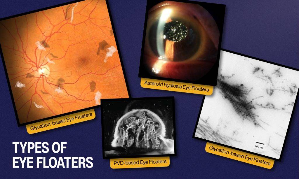
Eye floaters can meddle with your daily activities. Generally, the typical cause is the clumping of collagen fibers inside the vitreous body, the gel-like structure inside the eye. Glycation, a process in which the surface of collagen is oxidized and/or exposed to relatively high sugar levels, is linked as the cause. Subsequently, these clusters cast shadows on the retina and appear as squiggly lines, threadlike strands, cobwebs or rings.

However, there are several different types of eye floaters that we may experience throughout our lifetime. And with proper supplementation and nutrition, the condition is manageable, especially for collagen-based floaters. In order to help patients discern what they’re experiencing, we’ll look at these eye floaters and indicate which ones are manageable.

Embryonic Remnants

These types of eye floaters can form around the tenth week of pregnancy and occur when the hyaloid artery, which provides the eye with nutrients in the womb, pulls away from the lens. The process occasionally causes “debris” and is seen as floaters. These aren’t pieces of collagen from the vitreous but rather bits of the hyaloid artery and are not manageable with nutrition.

Asteroid Hyalosis

These types of eye floaters appear as white opacities in the vitreous, or stars shining in the night sky, and move around like typical floaters. They’re associated with age and are composed of calcium soaps, not collagen, and are not manageable with nutrition. Their cause is unknown.

Posterior Vitreous Detachment (PVD)

Floaters brought on by posterior vitreous detachment occur when the vitreous body physically pulls away from the posterior pole of the retina.  This process causes relatively large, collagen-based floaters to detach and release into the vitreous humor. 

What types of eye floaters can be managed?

The Floater Intervention Study (FLIES)1 showed that when the vitreous receives a continuous supply of antioxidant and enzymatic nutrients, the results showed a significant reduction in collagen-based floaters. These types of eye floaters are posterior vitreous detachment and glycation based.

The trusted scientists behind MacuHealth offer this patented nutrient blend under the name of VitreousHealth. It can be found online and through eye care professionals.

References

  1. Ankamah et al. 2021. doi.org/10.1167/tvst.10.12.19

Clinical Studies

macular degeneration causes
One way to find age-related macular degeneration causes is regular visits to your eye care professional.

Written by MacuHealth
Reviewed by Jim Stringham, Ph.D.

Currently, there is no cure for age-related macular degeneration (AMD). The eye disease affects over 11 million people and is one of the leading causes of blindness in the United States. But research shows that there are ways to prevent and manage the symptoms of this progressive eye disease.

Before we learn how to address and manage AMD, it’s vital to know its causes. And while the disease affects those over the age of 50, there are steps anyone at any age can take to avoid any possible future vision loss. We’ll look at macular degeneration causes and what steps you can take to manage this condition.

Dry Macular Degeneration Causes

According to WebMD, 85 to 90% of people with AMD have the early or “dry” form. When your eyecare professional conducts a dilated eye exam, they’re looking for drusen, which are accumulations of cholesterol and protein that collect under the retina. These small collections can limit typical eye operations. Researchers believe these clusters are caused by an inability of the retina to clean itself over time.

The existence of some drusen is normal and generally harmless. But as you age, drusen become harder and collects in the macula, which mediates central vision. As it accumulates, these particles keep oxygen and other vital nutrients from reaching your retina. You’ll notice a slow visual decline and, if left untreated, can progress to the more severe form of the disease, known as “wet” AMD. In this stage, small blood vessels in the eye begin to break and leak fluid beneath the retina.

Wet Macular Degeneration Causes

According to most researchers, drusen are also a wet macular degeneration cause. It happens when new blood vessels grow underneath the macula in an attempt to bring oxygen and nutrients to starved tissue. Unfortunately, these new vessels can break and begin to leak blood and other fluid into the eye, causing damage to your vision.

What Puts You at Risk For Macular Degeneration?

Researchers have determined several conditions and behaviors that can increase your chance of developing AMD. You’ll notice that some of these behaviors are habits you can stop or change.

Age. The disease is called age-related macular degeneration for a reason. According to WebMD, 2% of people in their 50s have AMD. Almost a third of people over the age of 75 do.

Family history of AMD. Researchers have discovered several genes linked to the disease. If someone in your family has had AMD, there’s a risk you could develop it as you grow older.

Race and ethnicity. Statistics show that Caucasians have the highest risk of developing AMD, with Chinese and Hispanic/Latinos following behind. African Americans are the least likely to have the disease.

Gender. Women live longer than men, which is why scientists believe two-thirds of those with AMD are female.

Smoking. Cigarette smoking is a macular degeneration cause that is preventable. Tobacco use reduces the amount of oxygen that reaches your eyes and produces an extremely high level of oxidative stress in the body. These factors can lead to the development of the disease.

Heart disease. A history of heart disease or high cholesterol could raise your risk of developing AMD.

Weight and diet. Some studies indicate that having a body mass index of over 30 can double your chances of getting AMD. And don’t forget that a vegetable-rich diet can decrease your chances of developing the disease.

What’s the Solution?

The most important thing you can do to prevent AMD is to see an optometrist regularly for an exam. Your eye care professional can offer sound strategies to help navigate your situation. And as shown above, quitting smoking, along with vigorous exercise and a balanced diet, go a long way in preventing and managing the disease’s symptoms.

Additionally, supplementation with nutrients known as carotenoids – specifically Lutein, Zeaxanthin and Meso-Zeaxanthin – can reduce the risk of developing AMD. Also, they can improve their visual performance. MacuHealth includes all three of the carotenoids that make up the macular pigment. The supplement is scientifically proven to protect the eye from damage, rebuild macular pigment and manage the symptoms of AMD.

Clinical Studies

What Causes Eye Floaters?
What causes eye floaters?

Written by MacuHealth
Reviewed by Jim Stringham, Ph.D.

It’s aggravating when oddly shaped particles move across your line of sight. These annoying strands are known as eye floaters. These pesky items can affect your ability to work, drive or go to the beach on a hot summer day. Reading a book, watching a movie or performing day-to-day tasks at home may develop into a struggle. It can even become difficult to concentrate when spending time with friends and family.

If floaters interfere with your everyday life, you’re not alone. The National Eye Institute predicts that nearly everyone will develop eye floaters at some point in their lives. One of the causes of eye floaters is a lack of nutrients in the part of the eye known as the vitreous body. For some, floaters are manageable and eventually go away.  But for others, they are a source of uneasiness and stress.

What can we do about these distracting spots and specks? We’ll take a closer look at what causes eye floaters and ways to manage them.

What is the Vitreous?

Before understanding what causes eye floaters, we need to learn about where they’re found: the vitreous. This clear gel-like substance consists of water, collagen and hyaluronic acid. It makes up nearly 80% of the eye and resides behind the lens and the front of the retina.

The vitreous helps maintain your eyeball’s shape, absorbs vibration and keeps the retina attached. It needs specific antioxidants to protect it against oxidative stress and disease.

Here’s What Causes Eye Floaters

If you’ve wondered what causes eye floaters, research shows the vitreous loses nutrients over time. This causes the collagen fibers inside it to clump together. These clumps, called floaters, cast shadows on the retina and become more noticeable when looking at the sun or bright surfaces.2 

If you’ve suffered from eye floaters for a long time, you’re likely familiar with how they float around (hence the name). Sometimes they’re black and gray colored dots. In some instances, floaters appear as squiggly lines, threadlike strands, cobwebs or rings. They can even emerge as a dark or light area of vision. Other times, they make some objects look blurry compared to the rest of your visual field. As your brain adapts to seeing them, you may notice them less, but they don’t truly go away.

Other Reasons for Eye Floaters

There is more than one answer to the question: What causes eye floaters? The symptoms generally become more common with age. But if you notice a sudden increase in the number of floaters, it’s time to see an eye care professional for an examination as it could be a sign of a more serious issue. One such condition is a detached retina. This happens when the vitreous separates from or peels off the retina, the part of your eye that processes light. It doesn’t cause your eye any pain, but it could lead to permanent vision loss if left untreated.

Other possible causes for an increase in floaters include eye inflammation, infection, retinal trauma, diabetic retinopathy, hemorrhaging or an eye tumor.

What Are the Treatments for Eye Floaters?

What can you do to maintain your eye health and manage floaters? Try eating more fruits and vegetables, wear protective eyeglasses (especially when it’s bright outdoors) and stay adequately hydrated. If you smoke, try to quit.

Your eye care professional may recommend risky, invasive therapies. These include using a laser to break up floaters. There is also a surgical procedure to remove the vitreous and replace it with a saline solution or bubble filled with gas or oil. This is known as a vitrectomy, and it comes with a risk of serious complications, including retinal detachment and cataracts.

Surgery may not be necessary, however.  Given that the vitreous requires a continuous supply of antioxidant and enzymatic nutrients, researchers from the Nutrition Research Centre Ireland hypothesized that giving participants these nutrients in a supplement, floater symptoms may subside. This trial, called the Floater Intervention Study (FLIES)1, showed a significant reduction in floaters and an improvement in visual function compared to placebo.

The trusted scientists behind MacuHealth are offering this patented nutrient blend under the name of VitreousHealth. This supplement is sold exclusively through eye care professionals.

References

  1. Ankamah et al. 2021. doi.org/10.1167/tvst.10.12.19

  2. Webb, Blake F et al. “Prevalence of vitreous floaters in a community sample of smartphone users.” International journal of ophthalmology vol. 6,3 402-5. 18 Jun. 2013, doi:10.3980/j.issn.2222-3959.2013.03.27

Clinical Studies

What are EPA and DHA
What are EPA and DHA? Read on!

Written by MacuHealth
Reviewed by Jim Stringham, Ph.D.

Omega-3 supplements are becoming more popular, especially those utilizing fish oil as an ingredient. Healthcare providers and optometrists are recommending patients increase their intake of this ingredient. According to recent studies, these polyunsaturated fatty acids are considered “good fats” because of their numerous benefits to the heart, brain and eyes. Yet many Americans fall short of the recommended intake of Omega-3s.

But when patients go to their local pharmacy or healthcare store, they see countless fish oil supplements on their shelves with many different formulas and price points. They’re confused about which one to choose. Many questions come to mind, such as: “What are EPA and DHA?” and “Which formula is the best?”

To help consumers make the best decision based on their health needs, we’ll look at the importance of certain ingredients in Omega-3 fish oil.

What to look for on an Omega-3 supplement label?

You might need to learn a whole new alphabet when looking at the label of a fish oil supplement. Be sure to check the letters in the active ingredients. You’ll find that many lower-cost bottles feature an Omega-6 called gamma-Linolenic acid, or GLA.

GLA can produce good results for the body as it has some anti-inflammatory properties. But it can also have potentially unhealthy consequences. The body can convert GLA into arachidonic acid, or ARA, which tends to be inflammatory. And because this ingredient is in poultry, eggs and red meat, Americans potentially have too much ARA in their diet. That can cause more inflammation, not less, leading to pain and swelling.

One more thing to look for when shopping for a high-quality fish oil product is if the base formulation is “re-esterified triglyceride” (rTG) or “ethyl ester” (EE). Re-esterified triglyceride is natural, recognized by the body and has high bioavailability. Ethyl ester is cheaper and easier to produce. But it has ethanol as its backbone, leading to health problems and lower bioavailability.

Then what letters should you be looking for on a fish oil supplement label? When it comes to reducing inflammation and dry eye symptoms, the research shows that EPA and DHA are the best. But what are EPA and DHA? We’ll explore what sets them apart from GLA below.

What are EPA and DHA?

According to SFGate.com, EPA and DHA are long-chain polyunsaturated fats with 14 or more carbons that have numerous health benefits. For example, your doctor may prescribe a steroid because of its anti-inflammatory properties. However, these drugs have serious side effects, including stomach pain, indigestion and an increased appetite. But the secret power of EPA is its ability to act like a steroid without the nasty side effects. EPA inhibits the enzyme that produces ARA, which can cause inflammation on the cellular level. 

It can be said that the more EPA you have in your system, the less harmful inflammation you’ll have in your body. It can ease the symptoms of arthritis, inflammatory bowel syndrome and other autoimmune disorders. EPA can also decrease the risk of obesity, heart disease, diabetes, depression, cancer and other diseases. 

DHA also has benefits for the brain and eyes and is critical to neural performance. It increases the speed of nerve signals, enhancing visual and cognitive performance and leading to improved reaction time, contrast sensitivity, and accuracy. And when EPA and DHA work together, it provides optimal benefits to the body.

Conclusion

We’ve answered the question: What are EPA and DHA? And despite the scientific evidence proving the importance of omega-3s to our vision and overall health, getting them into our diet is difficult. The body only receives these essential fatty acids through food such as fish, shellfish, algae or supplementation.

Taking a pharmaceutical-grade fish oil supplement delivers a safe and proven way to boost our vision, brain, and heart function. Because of evidence showing the benefits of taking EPA and DHA, MacuHealth developed TG Omega-3 Fish Oil to help patients meet current health demands.

TG Omega-3 is a natural re-esterified triglyceride formulation that is purified up to five times to eliminate toxins, impurities and fishy taste. TG Omega-3 offers 2,200 mg of EPA and DHA combined per serving. Next time you shop for fish oil, check the label to see if it has the correct formulation to give your body the boost it needs.